Showing posts with label lichens. Show all posts

Saturday, December 11, 2010

A lichen Malanelia subaurifera

I am an amatuer naturalist trying to learn something about everything living in my garden.

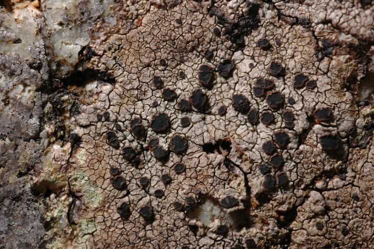
Photo 1 shows a lichen growing, amongst a number of similar patches, on a wooden bird table in my garden.

I am very far from being an expert on lichen identification but in the course of doing this blog over several years I have picked up a few tricks. One is to examine your lichen through a hand lens for any characteristic surface lumps on bumps. Some lichen species become decorated with powdery granules called soralia. Others with tiny sausage shaped protuberances called isidia. For my lichen, the latter are present in abundance as can be seen in the close-up Photo 2 (click on photos to enlarge).

Another trick to help with lichen identification is to check the under surface. In photo 2 I have peeled back a small section to reveal a black underside with a covering of tiny, root-like hairs. In the jargon, these are known as rhizines. Not all lichens have them. They are not roots in the traditional sense, since they do not function to suck-up water as do the roots of plants. Rather their job seems to be to help anchor lichens to surfaces.

Armed with the features above and my trusty copy of Lichens (Dobson, Richmond Publishing) I'm confident to identify my lichen as Melanelia (Parmelia) subaurifera. The book suggests a final test: gently rubbing the surface should leave a pale yellow-white abrasion. Photo 4 shows this.

I am fond of lichens and so was very pleased when someone recently made me a present of the new edition of Lichen Biology (Ed. Thomas H Nash III, publ. Cambridge). This book is primarily aimed at professionals and I don't pretend to have followed some of the very detailed sections on e.g. lichen biochemistry, but I was able to follow others and came away with a new respect for the intricacy with which nature adapts these little creatures to their environment. Take for example the construction of the little air-filled spaces often found inside the bodies of lichens:

Under a microscope a lichen is revealed to be a mass of long, spaghetti-like fungal cells ('hyphae'), mixed-through with a sprinkling of green, single-celled algae or sometimes cyanobacteria. (The fungus carries out various tasks such as water storage. The algae or cyanbacteria do what no fungus can: photosynthesise food from sunlight). In considering this description however it would be wrong to picture things as a random tangle of fungal threads and algal cells. Photo 5 shows a lichen cross section I made and discussed some time ago (here) and reveals that things are far from random. Of particular relevance for today's posting is the layer known as the medulla that contains a lot of air-filled voids (see the region of the box in photo 5 for example). What is the purpose of these voids? The answer of course is that algae, like all plants, 'feed' (via photosynthesis) on a diet of sunlight and gaseous carbon dioxide. This is the key to understanding the voids: they are present to allow the flow of gaseous CO2 gas to the algae.

So far so good, but possibly it might occur to you to wonder what happens when it rains?! Do these voids fill up with water and in so-doing stifle CO2 flow, and hence photosynthesis, in the lichen? As I learnt from the book above Nature, of course, has an answer. In one chapter, a remarkable photograph taken by Rosmarie Honeggar with an electron microscope reveals how the fungal threads in the medulla carefully coat themselves and their precious cargo of algal cells in a minuscule layer of water-repellent proteins. This remarkable water repellent 'jacket' prevents the medulla from becoming saturated with water and so maintains the algae in a gaseous environment conducive to photosynthesis.

The water-repelling proteins the fungal hyphae secrete are known as hydrophobins and their discussion would make a lengthy posting in its own right. They were unknown to science until the 1990's when they were discovered by Wessel and co-workers in a fungus called Schizophyllum commune. Since their discovery these remarkable molecules have turned out to be widespread amongst fungi, with fungi using them in a variety of ingenious ways to 'manipulate' the surface tension of watery environments. For example, in order to help their spores get airborne, some plant-infecting fungi coat their spores in hydrophobins so as to prevent them becoming trapped or stuck together by thin films of water. Hydrophobins also help the infectious spores stick to the waxy, water-repellent leaves of targeted host plants. You can find a short introduction and further references on hydrophobins in lichens here ( P.S. Dyer, New Phytologist (2002) 154 : 1–4).

So there you have it! Minuscule, air-filled voids in a wafer-thin lichen...but take a closer look and as so often in natural history, one finds a hitherto unimagined world of subtlety and sophistication.

Friday, September 17, 2010

A lichen: Lecidella elaeochroma

I am an amateur naturalist trying to discover everything that lives in my garden.

Once upon a time there was a large, upright apple tree in my garden. And then quite suddenly, one night, there wasn't!

What happened is a story most relevant for this blog...but one for another time. For today let me remark only that this calamity gave me an unprecedented opportunity to inspect my tree's upper branches for one of my favourite lifeforms - the lichens.

Photo 1 shows a lichen new for this blog (the grey smudge that is, the yellow is Xanthoria parientina I've blogged previously). Photo 2 shows a closeup of my lichen's areolate thallus (=cracked surface) and black, lecideine apothecia (= fruit bodies - see my artwork and explanation here).

Despite my fondness for lichens I am very far from being an expert. One problem facing the amateur is that many species look rather similar to the eye. Some are all but impossible to separate by anyone who does not happen to possess a forensic chemistry laboratory. (This is not a joke. It is not uncommon for the professionals to turn to e.g. chemical chromatography in pursuit of accurate identifications of lichens).

Fortunately there are a few 'tricks' available to the amateur. One is to observe your lichen under UV light. This is not as difficult as it sounds. In my case a battery-powered banknote reader from a 'pound store' (that's 'dollar shop', '100yen shop'... to those of you reading overseas) yielded the rather lovely result at the bottom of photo 2. The point is that had my lichen been the superficially similar Fuscidea cyathoides (picture available here), also occasionally found on wood, then UV light wouldn't produce the glow seen here. In the jargon, F. cyathoides is 'UV-' . By comparison, as I learnt from to my copy of Lichens (Dobson, Richmond Publ.) the common UK lichen Lecidella elaeochroma is 'UV+ orange' - clearly a good fit and my identification for today (as always I'm happy to be corrected).

I have mentioned previously a question that I have puzzled over regarding lichen: My (decidedly amateur) understanding of evolution has always been that, over time, it drives species towards adopting the optimal form for surviving in their environment. What has puzzled me is how then, it can be commonplace to see lichens with really quite dissimilar features occupying the same environmental niche. Inspect a few twigs on a tree and it's really not uncommon to find, side-by-side, both crustose (pancake-like) lichens, and, as here say, the bright yellow, foliose (=leafy) lichen X. parientina. To survive on wood, how can it be 'evolutionarily optimal' to be a bunch of bright yellow flakes, and optimal to be a grey pancake. Surely one ought to have 'won the argument'? It was satisfying recently therefore to come across a section in the book Introduction to Bryophytes (Vanderpooten and Goffinet, publ. Cambridge) that I think has given me the inkling of a solution to my confusion.

The book describes how some species of moss have become expert in seizing the opportuntity to colonise fleeting, virgin, environments. A newly appeared patch of burnt ground after a forest fire for example. Clearly being 'first moss on the scene' has the benefit you will enjoy the resources of your new home free from the pressure of competition. There is a price to pay for such a lifestyle however. To succeed at rapidly detecting newly emerged environments requires that you to put a great deal of your energies into sending out countless 'scouts' (a.k.a. spores) to explore your environs. By definition, if you're putting your energies into volume spore production, you are precisely not putting them into your own growth (producing lots of leaves etc.). Such 'fugitive mosses' therefore tend to be slight, quick-to-mature, normally annual plants, producing large numbers of small 20um spores.

Now 'fugitive' is not the only survival strategy amongst mosses. Enter the dominants. Dominants aim to out-compete other mosses for light and nutriants by growing faster and larger. This is a perfectly reasonable strategy, but again has its limitations. By investing a large proportion of their energy into the rapid growth of leaves etc. such mosses are left with little energy for the production of spores. Dominants then, will be less successful at rapidily discovering new areas, and tend to be larger, perennenial mosses with fewer spores. This is far from the complete story. As well as doimants and fugitives, the book above goes on to discuss the strategy of 'colonists', 'perennial stayers' and 'annual shuttles'. I have not found any freely available articles discussing similar issues for lichen (anyone?), but I think its not unreasoanble to suppose some similar ecology might hold for these fellow tree- and rock-dwellers.

All of which brings me back to my puzzle of how evolution can have a arrived at two such different forms (body shapes and colours) as optimal solutions for lichens living in the same place (a twig). I don't pretend a complete answer, but I feel I may have started to get an inkling of understanding. I think my confusion likely stems from woolly thinking on my part, namley, in erroniously imagining that evolution is about optimising a creature's form to fit a place. The more I've thought about the moss examples above however, the clearer it seems to me now that it is not 'body shape' that evolution is working to optimise, but rather the whole organism. That is, not merely its shape and colour, but rather the totality of its life cycle and survival strategy. Furthermore it is not sufficient to think of a lichen's 'environment' as being merely some unchanging point in space (the surface of a twig, say). This misses the very significant additional fact that our twig is subject to an annual cycle of dramatically changing seasons and that it itself is a growing, changing, transitory thing. At the risk of sounding too poetical, thinking of the lichens on my tree now I begin to get an image of a complex spaghetti of life histories and strategies at work. It is facile to try to ask whether 'yellow and flaky' is a 'better or worse' body shape for living on twigs than 'flat and grey'. Instead, each lichen will be following some complex survival strategy with multiple factors and tradeoffs. Viewed in this way, although two lichens have met on a twig in photo 1, viewed over extended time, the 'trajectory' of their lives is no doubt entirely different. The forms they have will be because these are the forms that best befit them to their individual, distinct, extended, life styles.

My great pleasure in researching this blog is that through it my view of my garden grows richer all the time. To my mind, no one has said it beter than Martin Luther

For in the true nature of things, if we rightly consider, every green tree is far more glorious than if it were made of gold and silver.

Sunday, February 21, 2010

A lichen Aspicilia contorta

I am an amatuer naturalist trying to learn something about everything living in my garden.

Returning to the iron manhole cover I discussed in my last posting, photo 1 shows yet another lichen eking out a living on the metal surface. From my copy of Lichens (F. Dobson, Richmond Publishing) I'm fairly confident the lichen here is Aspicilia contorta.

The little 'volcanoes' I've marked in the picture are the lichen's spore producing fruits (apothecia). If you're trying to identify a species of a lichen it's worth knowing that apothecia come in different 'flavours', in particular lecideine and lecanorine apothecia. This is well covered in the text books, but I wasn't able to readily locate a picture on the web so I've drawn one myself (I'm no botanical illustrator as you can see!). The difference is in the margin of the fruit body: The margins of lecanorine lichen apothecia tend to be the same colour as the main underlying lichen body (thallus) and contain algae. By contrst, the margins of fruits of lecideine lichens tend to appear distinct from the thallus and do not contain algae. The latter are often black (the books sometimes refer to the margin being 'carbonised'. I don't know whether this is just poor terminology or whether the lichen really does manufacture something akin to raw carbon in the margin. Anyone?).

From the description above it will be clear Aspicilia contorta is a lecanorine lichen.

Actually, my efforts to track down a description of all this on web led me to encounter a fair amount of technical jargon surrounding apothecia that had me quite confused at first. To save any fellow interested amateurs out there the trouble of going through the same, drawing two and the description below summarises the understanding I managed to pick up.

As I'd understood from my previous experiments slicing up apothecia, lichen fuits comprise an array of asci (= little tubes filled with spores). Photo 2 shows a single ascus I managed to separate from an apothecium I took from my garden A. contorta. As shown in drawing 2, the asci come sandwiched amidts a mass of upright fungal threads ('hyphae') known as paraphyses. The specialist textbooks often include pictures of these paraphyses, as they too can help in identifying the species of a lichen.

The fact that the asci are neatly arranged in an upright position is of course, no accident. It's important to the reproductive success of the lichen that when spores exit an ascus, they do so into the open air. There would clearly be no point in having the asci oriented haphardly and liberting a substantial proportion of their spores into back into the walls of the apothecium from where they've arisen in the first place. Of course, this only explains why the asci stand upright, it doesn't explain how they know which way is 'up'. I was fascinated to learn recently however (from a copy of the classic Spore Discharge in Landplants (Ingold) that I was lucky enough to find in a secondhand bookshop) that many cup fungi are phototropic (= sensitive to the direction of daylight), and use this to beneficially orient themselves. The book describes this for various cup-fungi, and I assume it is also the mechanism used by lichens, though strictly, I haven't come across a reference to confirm this (anyone?).

The asci + paraphyses are arranged on a thin layer known as the hypothecium. This comprises a mass of dikaryotic fungal cells, each containing two DNA nuclei. By a complex process of chromosome divison (mitosis) these cells eventually form (single nucleus-) spores. The whole business of sexual reproduction in lichens is complex and there is probably a lot left to discover. The pdf document here has some details.

The outer part of the fruit (technically termed the excipulum) comprises a mass of single-DNA-nucleus (haploid) fungal cells.

So now you know!

Today's has been a rather technical posting and perhaps marks a suitable point to leave my garden's lichens for a while (though I have yet to exhaust them). Outside the first signs of Spring are here: The volume of birdsong is increasing as the breeding season approaches and today on a country walk I spotted my first ladybird of the year (despite Oxfordshire still suffering regular frosts at night), all of which suggests I will not have to look too hard for new signs of life in my garden. Until next time...

Saturday, January 30, 2010

A lichen Lecanora dispersa

I am an amateur naturalist trying to discover everything living in my garden.

I have a manhole cover in my garden. (Try to contain your excitement at hearing this news dear reader!) This may seem an unpromising place to search for life, but a closer look reveals a host of those most enterprising (in terms of the habitats they are willing to conquer) of creatures - the lichens.

Zooming in, photo 1 (click on photos to enlarge) reveals at least two species: The yellow lichen (which looks like our old friend Caloplaca citrina - though I've not taken time to carefully check this) and a second species with numerous white, frosted fruits ('apothecia').

A little time spent with my trusty copy of Lichens (Frank Dobson, Richmond Publishing) and browsing some similar images on the internet, and I'm tolerably confident the latter is the lichen Lecanora dispersa.

There are at least 20 British species of Lecanora lichen. Dobson describes L.dispersa as 'very common and found even in the centres of cities'. It will grow on a variety of basic substrates including bark, stone, iron and leather.

Photo 3 shows a magnified view of the apothecia, with their white, wrinkled rims.

For photo 4 I took one apothecium and made a squash between a microscope slide and a coverslip. As I've discussed previously, lichens are a partnership between fungi and algae. The small green, algal cells are clearly seen in photo 4. That algae can be present throughout a lichen's cup-shaped apothecia was a new discovery for me - I'd hitherto only associated them with the non-fruit parts of a lichen.

The main purpose of a lichen's apothecia is to act as surfaces from which to liberate numerous reproductive spores. I've talked about this before, but I was not previously in a position to complement my descriptions with photographs. Recently however, I've been lucky enough to come into possession of a Cambridge rocking microtome. For those unfamiliar, this is a device for taking extremely thin (~few microns) slices of a specimen, which can then be viewed under a microscope. Actually things are not entirely straightforward, since, in order to avoid said slice ripping and disintegrating as it's cut, it's necessary to first provide support to the cellular structure of the sample by embedding the whole thing in a block of wax. The recipe for doing this is a little involved: Starting from pure water, you pass the sample through half-a- dozen water+alcohol baths. The baths are arranged so that the alcohol concentration steadily increases until finally your sample is left sitting in pure alcohol, the water in the sample's cells having, by then, been replaced with alcohol. Next, you substitute the alcohol for the solvent toluene (a solvent of wax) before dropping it into a bath of molten paraffin wax and leaving for several hours. Finally, you pour the wax+sample into a mould and leave to cool. (In fact, there are still futher steps that finesse this process - but you get the general idea! Should you wish to try it yourself the best guide I've found is the booklet Practical Microscopy (Eric Marson, Northern Biological Supplies) and/or you could join one of the hobbyist microscopical societies such as the redoubtable Postal Microscopical Society.)

Photos 5 and 6 show my rocking microtome. The blue handled blade is the extremely sharp sectioning razor. The white object is the wax-block-embedded sample. Pulling the lever at the rear of the microtome (photo 5) raises the sample and releasing (photo 6) forces it down onto the blade shaving off a wafer thin slice and simultaneously ratcheting the sample forward ready to take the next.

And the consequence of all this labor?... Photo 7 shows a cross section through a lichen apothecium. Strictly, for this posting it ought to be that of L.dispersa, but working on the principle 'walk before you can run!' I instead sectioned a sample of our old friend X.parietina from my apple tree, since the latter has large apoethecia and a robust surrounding thallus (= 'body'). I've labelled up photo 7 (click to enlarge) and you can clearly see that the apothecium surface (technically termed the hymenium) comprises an assemblage of sausage-shaped 'asci' packed full of spores.

One thing puzzles me: I have not seen the granular bodies I see here labelled up in the handful of books I have on lichen. Possibly these are air bubbles. They turned up very frequently in my samples however and I don't believe they are. Can anyone comment?

Tuesday, March 24, 2009

A black lichen Placynthium nigrum

I am an amateur naturalist trying to discover everything living in my garden.

Spring is well and truly springing here in Oxfordshire. Soon animals and plants will be appearing in my garden faster than I can photograph them, let alone write about them. Whilst things are still moderately calm therefore, I'm seizing the moment to press on with my task of cataloguing my garden's lichens.

Quietly going about its business, photo 1 (click on photos to enlage) shows a black crustose lichen (for the uninitiated, see my explanation here) that decorates my garden wall in places. Photo 2 is a closeup (I've slightly digitally sharpened this image using software).

I'm no expert, and happy to be corrected, but from what I can tell from leafing through my copy of Lichens (Frank Doson), although there are numerous lichens with black fruiting bodies ('apothecia') on otherwise coloured 'backgrounds' ('thalli'), there are only a handful of mostly- or wholly-black crustose lichens to be found in Britain. A number are marine. Verrucaria maura, for example, is common on rocky shorelines where it is somtimes mistaken for oil pollution.

Of the mostly black, 'land-locked', lichens, I spent some time looking at the photos of Verrucaria nigrescens on the excellent 'UK Lichens' site. Looking closely however, this seems to have a more chocolate-brown thallus, albeit one peppered with many black 'perithecia' (see my definition here).

On balance therefore I'm tentatively identifying my lichen as Placynthium nigrum which my copy of Dobson describes as being "Very common, mainly on hard calcareous substrates throughout Britain. Often found on flat tombstones and cement".

A slight puzzle is that the photo in Dobson shows a more powdery ('coralloid') surface than is evident in my photo 2, although the book adds this lichen may be "sometimes smooth and cracked especially in polluted areas". Where I live is rural and I don't believe especially polluted. That said, some lichens are extraordinarily sensitive to even minute amounts of air pollution - whole books have been written on this topic. Anyhow this variability in texture gives me some added confidence in my identification.

Turning to my copy of Lichens (Oliver Gilbert, New Naturalist Library) a nice thing to discover was some growth-rate data for Placynthium nigrum. I learn that young patches expand their radius at 1.66mm/year and mature patches at 0.08mm/year. Lichenometry is the technique of dating old structures (churches, stone circles etc.) by studying their lichen populations - you can find an article here. Applying the data above to the approximately circular, 10mm-radius, patch in photo 2 ages my lichen at between 6- and 125-years old! Not the most exact figure I grant you, but fun to know.

As I commented in a recent posting, I am puzzled by the colours lichens adopt. Over the millenia animals have been driven to evolve their numerously-coloured fur coats, feathers and exoskeletons so as to optimally attract mates, hide from predators, advertise their venomous stings etc. Similarly my amateur's understanding is that plants are mostly green by virtue of the need to pack their leaves and stems with chlorophyll. Even various of the larger mushrooms have evolved specific colours, presumably to alert browsers to their poisonous nature or advertise their presence to 'pollinating' (botanists may wish to turn away at this point!) insects. Some even glow in the dark for this very purpose. But how it is that some lichens on my garden stonework gain advanatage by being coloured matt black, whilst others 'prefer' greyish/white and still others, bright yellow, I struggle to guess. Can anyone help?

Today's posting brings my garden lichen species-count to eight. I feels that I may be approaching completion with regard to this particular lifeform. Until, that is, I find another dozen species through more careful inspection of my garden's rocks and trees. Stood outside earlier today for example, when acquiring the photos above I was aware only of our black friend and of the grey-white patches which (though I haven't checked in detail) I'm assuming is our old friend Verrucaria. Sitting now at my computer screen however, staring at an enlarged version of the photo 1, I'm suddenly noticing the array of tiny orange blobs towards the centre of the image. Time to go back outside methinks!

Sunday, February 8, 2009

A lichen Aspicilia calcarea

I am an amatuer naturalist trying to discover what lives in my garden.

Those following my recent posts will know I have been on something of a mission to blog the lichen-life on the exterior of my house. Photo 1 shows yet another inhabitant - another crustose lichen (for those unfamilar with lichens, see my post here).

Incidentally, photo 1 also captures (upper left) our old friend, the moss, Tortula muralis.

After a little research I'm confident the lichen here is Aspicilia calcarea. Characteristic features include the cracked, white thallus (the main body of the lichen) and the irregularly shaped apothecia (the black, spore-liberating cups) sunk into the thallus. The books tell me that A. calcarea is common on hard calcareous walls etc. in lowland Britain. Photo 2 shows a closeup.

For those wanting a cheap photographic key to some common, British, urban lichens incidentally, I recommend the short-form guide sold by the good people of the Field Studies Council . For something more detailed the book Lichens (Frank S.Dobson) is especially good.

I'm fond of lichens. Their ability to shrug off the worst the elements can throw at them gives them, for me, an appealing minature 'feistiness' - I picture them squatting on exposed boulders on windswept mountain sides goading the rain "Come on! Give me your best shot! Is that all you've got pal !?"

On a more rational note (!), something that intrigues me is the diverse array of colours and shapes lichens adopt. I have no deep expertise in evolutionary ecology but as I understand it, there is nothing haphazard about the forms taken by species. Life is hard and an ever-present scarcity of resources and the threat of predation and disease is a constant imperative, forcing species to individually specialise in unique methods of suvival. A famous example is of course the beaks of finches, with different species having been driven to evolve different beak-shapes to allow them to eat different nuts and seeds. Different birds evolving different beaks to help them occupy different feeding niches is one thing. The distinct environmental pressures or purposes that drive two lichens such as A. calcerea and C. citrina to adopt such different colours and (once you look closely) really quite different textural forms, when both seemingly occupy the same ecological niche of lowland stone (indeed, the same household windowsill in my case!) - I struggle to guess. Do leave a comment if you can help me.

Saturday, January 17, 2009

A lichen Caloplaca citrina

I am an amateur naturalist trying to discover everything living in my garden.

I've previously blogged the yellow lichen Xanthoria parietina that grows on the upper, east-facing exterior window sills of my house. On the lower sills there's another yellow algal/fungal partnership taking place (photo 1 - click to enlarge), this time in the form of a crustose, Caloplaca lichen.

I'm not a lichen expert, but having referred to the text books, I'm fairly confident the species here is Caloplaca citrina. The apothecia (the little, yellow spore-producing 'pin cushions') are spread about over a powdery, yellow thallus. Were this C. holocarpa (another common Calopaca with yellow/orange apothecia) the thallus would be grey. The thallus of C. dalmatia is cracked by thin black lines. The lack of any change in lichen texture / colour near the perimeter of the patch also distinguishes it from other various other superficially similar Caloplaca species such as C. decipiens. Identifying lichen is all about noting the tiny details!

There is yet another common possibility: the superficially similar powdery, yellow lichen Candelariella vitellina. In this case however there is an acid test (if you'll forgive the (chemically imprecise!) pun) to tell the two apart: a tiny drop of potassium hydroxide applied to most Caloplacas will turn them red. Candelariellas on the other hand, show no reaction. Photo 2 shows the positive result in my case.

I've read a suggestion that the purpose of yellow colouration in lichens is to provide protection against the harmful UV part of sunlight. I'm happy to accept this but it makes me wonder why only some lichens need to bother (there are plenty that don't: green, grey, white and even black lichens being commonplace - the two Verrucaria lichens I've previously blogged for example) .

Finally, the fact that C. citrina occupies the lower sills or my house and X. parietina the upper ones, makes me wonder whether the latter is more tolerant of low light conditions. Can anyone tell me?

Monday, February 11, 2008

Two Verrucaria lichens

I am an amateur naturalist attempting to identify all the life in my garden.

February is seeing Oxford hit by a succession of clear, frosty days. The cold weather means that many of my garden's plants, fungi and animals are tucked away out of sight or sitting out the winter in a dormant state. As I've remarked before, this is a good time for the amateur to take an interest in those humble lifeforms the mosses and lichens which, far from lying dormant, seem to positively relish the winter and the lack of competition it affords. My garden wall for example, is currently layered with a vibrant mat of fruiting mosses.
Photo 1 shows a closeup of two more denizens of my wall's stonework. For the uninitiated, no, this is not a mistake! What appears from a distance to be little than a collection of stains on the stonework is in fact my blog's first example of a crustose (=flat, crustlike - see here for more explanation) lichen. In fact there are two in photo 1. Photo 2 shows a zoomed-out image of the same region. Personally, I find it little short of miraculous to see life thriving on bare rock.

Photo 3 shows a 40x magnified view of the tiny black dots peppering the surface of the uppermost lichen in photo 1. These are perithecia - tiny flask shaped 'salt cellars' from which the lichen shoots out spores.

When it comes to species, as always, my identifications come with a health warning: I'm far from expert in identifying lichens and furthermore, without going to the lengths of studying spores under the microscope and applying chemical tests (lichenologists take these seriously: I even have a book - Microchemical Methods for the Identification of Lichens, Orange, James and White - that teaches the amateur how to perform chromatography!), it can be well-nigh impossible to definitively identify some lichens by visual appearance alone. With this disclaimer, let me say that my lichen's fruit bodies being set in little craters in the surface, and the black line delineating the thallus perimeter in photo 1, are both features that fit with the description of Verrucaria baldensis in my copy of Lichens:An Illustrated Guide to the British and Irish Species (F.S.Dobson).

A similar analysis for the lower, 'less spotty' (though you'll still see some perithecia if you look closely) lichen in photo 1 inclines me to an identification as Verrucaria muralis.

The Verrucaria genus of lichens is noteworthy for including a number of freshwater and maritime lichens. The next time you're at the seashore, look towards the high-tide line and in all likelihood you find the rocks everywhere stained black. This isn't oil (as is sometimes thought) but a result of the ubiquitous seashore lichen V.maura.

Amongst the many extreme features of the life of a crustose lichen is the extraordinarily slow growth of some. According to my copy of Lichens (O. Gilbert, New Naturalist series) lichens are amongst the oldest and slowest growing organisms in the world: Rhizocarpon geographicum, for example, puts on a mere 0.09mm of growth-radius per year. A patch of the lichen Aspicilia calcerea, growing on the Rollright Stones, a stone circle in Oxfordshire, is estimated to have begun life there in 1195AD. It's thought some Icelandic lichens might be 9000 years old! I'll have nothing quite so extreme in my garden, but I am curious to track the growth rates of at least a few of the crustose patches on my garden wall. Watch this space!

Tuesday, January 29, 2008

A lichen Physcia tenella

I am an amateur trying to learn something about everything living in my garden

I promise to return to more sizable and familiar lifeforms soon, dear readers! For today's posting however: another lichen.

The lichen seen in photo 1 (click to enlarge) decorates many of the branches of my garden apple tree (at (1.2,1.5) - see here) and I'm almost certain (see shortly) in identifying it as Physcia tenella ('it' being either of the two greyish patches below the coin, as opposed to the yellow lichen left of the coin which - though I've not examined it is detail - is probably our old friend X. parietina)

The meaning of the Greek physcia (pronounced 'Fisk-ee-a') seems to be a matter of debate on the internet. I have come across it variously translated as "bellows", "breath-like", "sausage-shaped" and "blister"! Can anyone clarify the true meaning?

P.tenella is my blog's third example of a foliose lichen. According to my copy of the scholarly Lichens (F.S. Dobson, The Richmond Publishing Co.), Physcia lichens can be distinguished from other foliose lichens, in part, by their narrow (<~2mm) lobes, and septate spores (septa being thin, internal dividing walls - see here for some examples) - though I confess I've not looked for the latter.

Two further distinguishing features of P.tenella are i) the presence of eyelash-like cilia on the edges of the lobes. You can see these in the 40x-magnified photo 2. (Can anyone tell me what purpose it serves to lichen to 'sprout' these?) And ii) the presence of granular soredia (see here for my explanation of what these are) decorating the ends of the lobes. Were I to observe the soredia 'packaged' together inside little 'hoods' I understand I'd be looking at the closely similar lichen P.adscendens (since its not obvious they are, I'm sticking with my identification as P. tenella).

Finally, two nice facts about P.tenella I got from reading my copy of Lichens (O.Gilbert, The New Naturalist series). Firstly, P.tenella is one of three British lichen species favoured by long-tailed tits (Aegithalos caudatus) in building their nests (on average each nest contains nearly 3000 lichen flakes!). Secondly, being fairly acid- and nitrogen-tolerant (not all lichens are), in areas where people walk dogs, P.tenella is one of the commonest lichens to colonise the region marginal to that patch of tree-trunk known to professionals as "the canine zone" . It shouldn't be hard to imagine what this is!

Tuesday, January 1, 2008

A lichen Punctelia (Parmelia) subrudecta

I am an amateur naturalist trying to work out all the things living in my garden.

What better way than to start 2008 than with my second lichen (photo 1 - click to enlarge).

This one is growing on a bough of my garden apple tree (at (1.2,1.5) - see here) and is once again an example of a foliose (flakey, 'leaf like') lichen.

As explained in my previous posting, lichens are an organism that comprise a combination of a fungus and an algae. You might expect a curious life form such as this to have some unusual means of propagation and dispersal. In fact, lichens have at least three. The method(s) a given lichen favours can be a handy hint towards identification.

One method by which lichens disperse, as discussed last time, is via spores produced from fruit-like bodies called apothecia. The white bumps peppering my lichen's surface (thallus) in photo 1 are a second. As photo 2 (40x magnification) shows, these bumps consist of small white granules known as soredia. Soredia are little lumps of lichen (some fungal hyphae and a few algal cells) whose purpose is to flake away, hopefully to land somewhere new where they can set up home.

With regard to reaching a species identification, as I've said before I'm basically a novice when it comes to lichens. Based on the very handy A Key to Lichens on Twigs (Wolseley, James, Alexander) - a leaflet produced by the excellent Field Studies Council, together with a copy of the highly scholarly Lichens (F.S. Dobson, Richmond Publishing Co.) that Santa was generous to deliver me recently, I'm going with the identification Punctelia (Parmelia) subrudecta. I'm not 100% confident in this as the books above describe this species as having a grey thallus, whereas I'd definitely say that in my photo is greenish. That said however, the photo of P. subrudecta on the excellent site of the Botanical Museum of Oslo seems a good match. Furthermore, as I mentioned in my previous lichen posting, another handy tip for the identification of lichens is the use of chemical tests: according to the books, P. subrudecta turns red when exposed to a drop of sodium hypochlorite (=household bleach). The reaction was small and fleeting, but as shown in photo 3 (you'll want to click to enlarge), removing a small piece of my lichen and testing it resulted in just such a reaction. In short, I'll stick with identification for now and inmvite the experts out there to correct me!

Monday, October 1, 2007

A Lichen Xanthoria parietina

I am an amateur naturalist trying to discover a little about everything living in my garden.
Quietly minding its own business on an exterior concrete windowsill of my house is the lichen shown in photo 1 (click on photos to enlarge). It also grows on the branches of the apple tree in my garden.

My knowledge of the 1854 species of British lichen is extremely limited, but this one, Xanthoria parietina is so common in the UK that it is one I do happen to know.

So what is a lichen? As I discovered recently from reading Lichens (Oliver Gilbert, Collins New Naturalist) this was a question that caused much heated debate amongst 19th century naturalists, with some claiming lichens were plants and others fungi. It turns out that both parties were right: lichen comprise a fungus working "in harmony" with algae (or, in about 10% of cases, with cyanobacteria). The fungus makes up most of the lichen body and is the "stuff" you see when you look at a one. The fungus also takes care of a lot of the tasks associated with water and food storage. The algae (or cynaobacteria) perform photosynthesis i.e. turn sunlight into food. A quote attributed to one Trevor Goward, I came across on this site sums things up nicely:

"Lichens are fungi that have discovered agriculture"

Under the microscope, a cross sectional slice (acquired after no small amount of effort fiddling around with a razor blade at my kitchen table) of my lichen shows things up clearly. Photo 2 (400x, click to enlarge) shows the layer of green algal cells (which I read are algae of the genus Trebouxia in the case of X.parientina) sandwiched between layers of fungus (the various layers, taken from top to bottom, being termed the upper cortex; medulla; and the lower cortex).

So, how does one set about identifying a lichen? Firstly, it's important to know that lichen fall into three main categories: crustose these being the lichens that form patches of negligible thickness like patches of gum on a city pavement; foliose, lichens composed of more visible "flakes" (like the X.parietina in photo 1); and fruticose - little hairy/fibrous "clumpy" lichens that mostly frequent trees.

Next, many lichen are selective about the surface on which they will grow: In particular, some require an acidic surface others alkaline. Putting a drop of lemon juice (=an acid) onto a surface and examining it through a hand lens, if the surface is alkaline small bubbles will be seen forming in the droplet.

A next trick widely used by lichenologists to aid identification makes use of chemical colour tests. Applying certain chemicals (such as household bleach and potassium hydroxide solution) to certain lichens results in powerful colour changes. X. parietina turns bright scarlet in response to 10% KOH-solution for example. As I learn from Dr. Gilbert's book, the complex colouring of lichens results from the presence of acidic crystals that lichens secrete within their body. It seems no one really knows the purpose of these compounds, but over 400 types have been discovered.

Finally, for the experts, examining a lichen under a microscope can be a strong aid to identification. Assuming some are present, a particularly revealing part of a lichen to examine is an apothecium. Apothecia are the little circular cup-shaped fruiting bodies visible in photo 1. Photo 3 shows a 1000x magnified view of a small section of one of them and reveals it as comprised of numerous asci - little tubes full of spores. The asci eventually burst open, liberating the spores and so ensuring reproduction. It turns out that in response to certain iodine-based solutions (I've used Melzers agent in photo 3) the tips of the asci of different lichen species stain in characteristically different ways (clearly, staining blue in photo 3). The classic book to turn to for details is Lichens:An Illustrated Guide to the British and Irish Species (F.S.Dobson).

This discussion barely scrapes the surface of the fascinating natural history of lichens: there is their extraordinarily slow growth and the great age some attain; the fact that sporulation is only one amongst a variety of ways that lichen have of reproducing; the extreme sensitivity of certain species to air bourne pollution; and the relationships lichens have with other plants and animals. Sadly, space means a discussion of any of these will have to wait for a future posting. Instead, I'll leave you with a quote from Dr. Gilbert's book attributed to A.C.Benson 1904

"Beauty, beauty? What is it? Is it only a trick of old stone and lichen in sunlight?"

I'll have a look at my windowsill and let you know!